已添加
登录/注册
变成可以直接使用的白底素材
变成可以直接使用的白底素材
金国桢
2026.04.15
编辑
变成可以直接使用的白底素材
金国桢
变成可以直接使用的白底素材
金国桢
牙齿釉质结构科学示意图,清晰展示牙齿剖面层次,用箭头标注氟元素加固牙釉质的过程,示意图侧边放置一个防护盾牌图标,整体风格简约专业、易理解
1q08y1boi8-HB
医学横截面、超写实、牙齿剖面结构清晰(牙釉质/牙本质/牙髓分层)、专业医学可视化,生成口腔牙体横截面超写实医学CG图像:同一磨牙纵切面,根管内牙胶尖填充完好,牙冠部窝洞已分层充填复合树脂材料(牙齿色,半透明质感),树脂与牙体组织密合,咬合面形态已恢复牙齿正常解剖外形(包括窝沟点隙)。牙髓腔已被树脂封闭,与外界隔绝。牙齿整体形态完整,外观与正常牙齿一致。超写实医学可视化,高清,树脂与牙体界限隐约可见。
GD256518266
科学示意图风格,展示牙齿被酸逐渐腐蚀钻洞的过程,清晰呈现牙齿剖面结构和龋洞形成,标注简化易懂,适合科普讲解
GD183351987
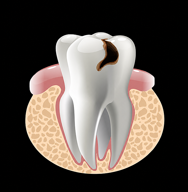
参考图片中龋齿牙齿剖面解剖示意图风格,更换为黑色背景
rainbownn
分子结构示意图,清晰展示丙烯酸分子通过化学铰链基团连接到淀粉分子的羟基上的化学键结构,画面为科学插画风格,背景为纯白色,分子结构标注清晰,配色简洁专业,符合化学学术绘图规范
GD358475498
参考原图风格,重新生成补牙过程的医学科普插画,保持六个步骤的布局和文字说明:龋齿、去腐备洞、充填、光固化、调合抛光、治疗完成,浅蓝背景,清晰扁平医疗插画风格
用户sma3FIKE
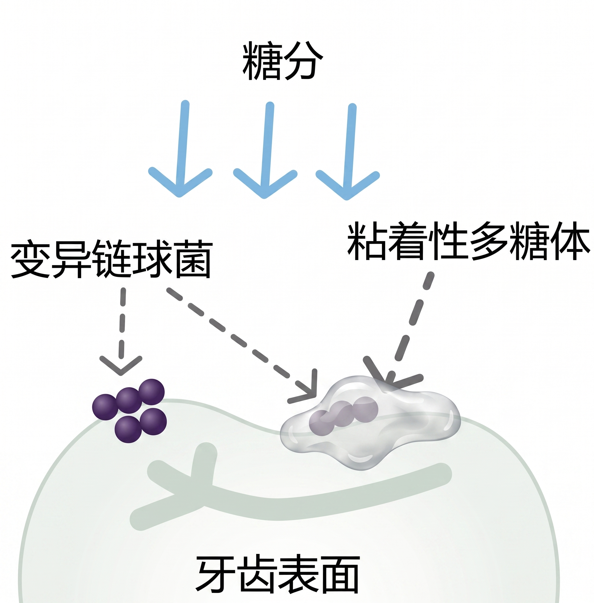
参考提供的图片风格:卡通可爱手绘风格,蓝白清新配色,牙齿拟人化设计,文字排版层次分明,分三个步骤用箭头连接,每一步配拟人化小插图,内容分别是:第一步糖+细菌=酸,第二步酸腐蚀牙齿,主标题文字"3步看懂:糖怎么‘吃’掉牙齿",正文文案:第1步:你吃了糖,糖留在牙齿表面;第2步:嘴巴里的细菌吃了糖,排出"酸";第3步:酸慢慢腐蚀牙釉质,形成蛀牙;总结文字:简单说:糖本身不伤牙,细菌吃了糖产的酸才伤牙。
1q5jd14daf2drc88-HB
牙齿
GD548874114
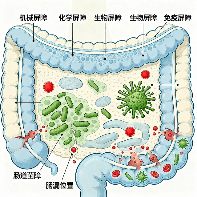
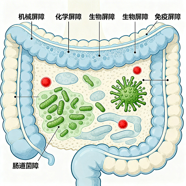
医学科普插画展示人体肠道的四大屏障,清晰标注四个屏障分别为:机械屏障、化学屏障、生物屏障、免疫屏障,结构清晰,科学准确,风格专业易懂,配色舒适
67brgu3ls-HB
统一背景色
GD512676845
消除
GD512676845
医学横截面、超写实、牙齿剖面结构清晰(牙釉质/牙本质/牙髓分层)、专业医学可视化,生成口腔牙体横截面超写实医学CG图像:同一磨牙纵切面,根管内牙胶尖填充完好,牙冠部窝洞已分层充填复合树脂材料(牙齿色,半透明质感),树脂与牙体组织密合,咬合面形态已恢复牙齿正常解剖外形(包括窝沟点隙)。牙髓腔已被树脂封闭,与外界隔绝。牙齿整体形态完整,外观与正常牙齿一致。超写实医学可视化,高清,树脂与牙体界限隐约可见。
GD469713083
生成“降低敏感反应”的示意图
Loku0725
1q08y1boi8-HB
[主图 2] 漱口水核心功效成分解析图。[氛围/风格] 专业科研医疗级氛围,干净通透,光线均匀柔和,商业精修质感。[元素/光影] 全屏展示高精度3D透明抑菌分子和修护分子结构,错落排版,用纤细浅灰色细线标注每个成分对应的功效:抑菌分子标注“长效抑菌”,修护分子标注“修护口腔”。分子结构透明轻盈,背景为浅灰白色渐变,保持整体干净专业。商品主体占比控制在60%-80%,画面满铺无边框。[配色色系] 主色浅灰白,透明分子淡蓝色和淡绿色,标注文字深灰色,整体配色干净专业。[视角构图] 正中心构图,整体画面均衡,景深适中。[文字版式] 每个成分用细线连接对应的功效文字,文字小号左对齐,深灰色清晰易读,不遮挡主体结构。主标题“核心功效”在顶部居中,大号粗体深灰色。[比例分辨率] 3:4比例,2k分辨率;文字清晰锐利,画面超高清。[负向提示词] 变形,水印,文字错乱,背景杂乱,色彩暗沉,结构模糊。
1q0jx33yjk-HB
医学科普肠漏示意图,展示肠道屏障受损后,毒素和有害物质穿过肠壁进入血液的病理过程,结构清晰,科学准确,和原图保持一致的插画风格,标注清楚肠漏位置
67brgu3ls-HB
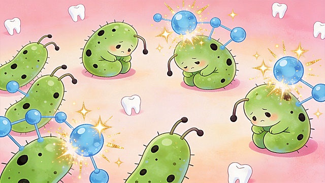
可爱卡通儿童插画,一群蛀牙细菌被氟分子击退,细菌变得无精打采,画面色彩明亮、风格软萌可爱,适合小朋友观看
1q08y1boi8-HB
修改这张医学科普口腔问题信息图,将图片中标题“引发口腔‘连锁反应’”更换为更粗的现代无衬线字体,配色改为深蓝色渐变
涵博2025
一颗细菌的剖面示意图,左右两半完全对称但结构截然不同;左半为紫色厚壁,壁层致密如树轮,内部锁满深紫色颗粒状结晶紫-碘复合物;右半为红色薄壁,壁层疏松可见孔隙,紫色颗粒正从孔隙中向外流失滴落;中央竖直线分割左右,底部一行小字“结构决定结果”;极简矢量图标风,白底,线条干净清晰,适合PPT图标使用。
GD220794368
牙齿涂氟前后对比示意图,左侧一颗带有早期白斑的牙齿标注文字"早期蛀牙",右侧是涂氟再矿化后恢复光滑的牙齿,用箭头连接并标注文字"修复中",清晰直观展示修复效果
1q08y1boi8-HB
close up of water flosser removing food debris between teeth, water jet, dental care, clean teeth, educational diagram style
GD790361524
给我生成一个类似附件的细胞图片
1qk2bwi75d6Lm0yw-HB
科研示意图 人溶菌酶在人体免疫细胞分泌的机理简图,清晰简洁的科学插图风格
GD113876734










![[主图 2] 漱口水核心功效成分解析图。[氛围/风格] 专业科研医疗级氛围,干净通透,光线均匀柔和,商业精修质感。[元素/光影] 全屏展示高精度3D透明抑菌分子和修护分子结构,错落排版,用纤细浅灰色细线标注每个成分对应的功效:抑菌分子标注“长效抑菌”,修护分子标注“修护口腔”。分子结构透明轻盈,背景为浅灰白色渐变,保持整体干净专业。商品主体占比控制在60%-80%,画面满铺无边框。[配色色系] 主色浅灰白,透明分子淡蓝色和淡绿色,标注文字深灰色,整体配色干净专业。[视角构图] 正中心构图,整体画面均衡,景深适中。[文字版式] 每个成分用细线连接对应的功效文字,文字小号左对齐,深灰色清晰易读,不遮挡主体结构。主标题“核心功效”在顶部居中,大号粗体深灰色。[比例分辨率] 3:4比例,2k分辨率;文字清晰锐利,画面超高清。[负向提示词] 变形,水印,文字错乱,背景杂乱,色彩暗沉,结构模糊。](https://gd-cms.dancf.com/gd-cms/xi19e5/0/5e628348-27ea-49fb-8cc3-5ea0c5c6dbed21637.png?x-oss-process=image/resize,w_640,type_6/sharpen,120/interlace,1)