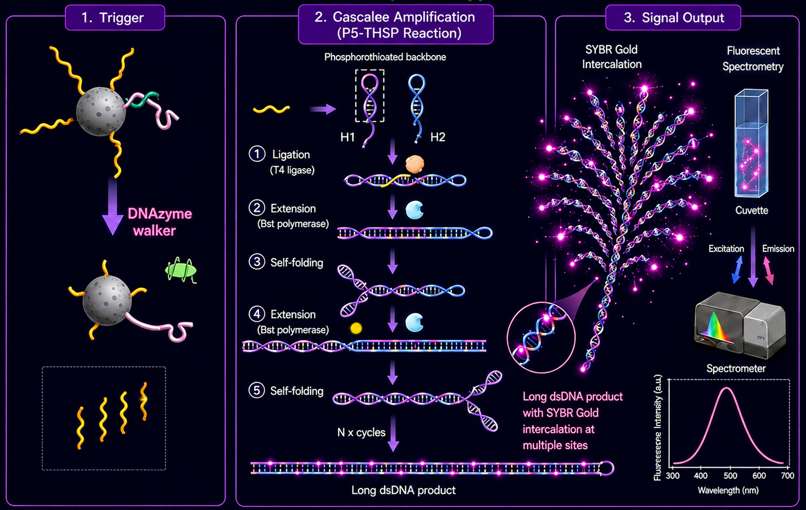

优化这张DNA生物机制示意图的绘制细节,保持整体设计、布局、文… - 图片创意预览

优化这张DNA生物机制示意图的绘制细节,保持整体设计、布局、文…

优化这张DNA生物机制示意图的绘制细节,保持整体设计、布局、文字内容、颜色风格完全不变,不要重画内容,不要改变任何元素的位置和形状。仅优化所有DNA双螺旋线条,使其连贯流畅,没有断开、接口或不连续的接

用户头像

1rax2bq7r6E...

2026.09.10

Ultra-detailed scientific illustration, flat vector style, showcasing the process of "Triparental Mating" for bacterial plasmid transfer. The image is divided into three clear stages from left to right. Stage 1: Three distinct bacterial cells in close proximity. 1 Donor E. coli: Contains a "mobilizable plasmid" with a visible "gene of interest" (like a fluorescent protein or antibiotic resistance marker) and an "oriT" site. 2 Helper E. coli: Contains a "conjugative plasmid" with visible "tra genes" (transfer genes) and pilus structures. 3 Recipient bacterium (e.g., a Rhizobium cell): Has a different cell wall shape, labeled, with no plasmid inside. Stage 2: The helper E. coli extends a "pilus" (a thin tube) to attach to the donor E. coli. The conjugative plasmid's tra genes mobilize the mobilizable plasmid. Show a clear arrow indicating the transfer of the mobilizable plasmid FROM the donor TO the helper cell. Stage 3: The helper E. coli, now carrying the mobilizable plasmid, extends a new pilus to attach to the recipient bacterium. Show a second arrow indicating the final transfer of the mobilizable plasmid INTO the recipient. The recipient bacterium now contains the plasmid. Use a distinct, consistent color scheme: Donor is Blue, Helper is Orange, Recipient is Green, Mobilizable Plasmid is Red, Conjugative Plasmid is Yellow. The background is clean white. All key elements have clear labels with leader lines. The style is educational, schematic, and extremely clear for a presentation slide.Ultra-detailed scientific illustration, flat vector style, showcasing the process of "Triparental Mating" for bacterial plasmid transfer. The image is divided into three clear stages from left to right. Stage 1: Three distinct bacterial cells in close proximity. 1 Donor E. coli: Contains a "mobilizable plasmid" with a visible "gene of interest" (like a fluorescent protein or antibiotic resistance marker) and an "oriT" site. 2 Helper E. coli: Contains a "conjugative plasmid" with visible "tra genes" (transfer genes) and pilus structures. 3 Recipient bacterium (e.g., a Rhizobium cell): Has a different cell wall shape, labeled, with no plasmid inside. Stage 2: The helper E. coli extends a "pilus" (a thin tube) to attach to the donor E. coli. The conjugative plasmid's tra genes mobilize the mobilizable plasmid. Show a clear arrow indicating the transfer of the mobilizable plasmid FROM the donor TO the helper cell. Stage 3: The helper E. coli, now carrying the mobilizable plasmid, extends a new pilus to attach to the recipient bacterium. Show a second arrow indicating the final transfer of the mobilizable plasmid INTO the recipient. The recipient bacterium now contains the plasmid. Use a distinct, consistent color scheme: Donor is Blue, Helper is Orange, Recipient is Green, Mobilizable Plasmid is Red, Conjugative Plasmid is Yellow. The background is clean white. All key elements have clear labels with leader lines. The style is educational, schematic, and extremely clear for a presentation slide.