已添加
登录/注册
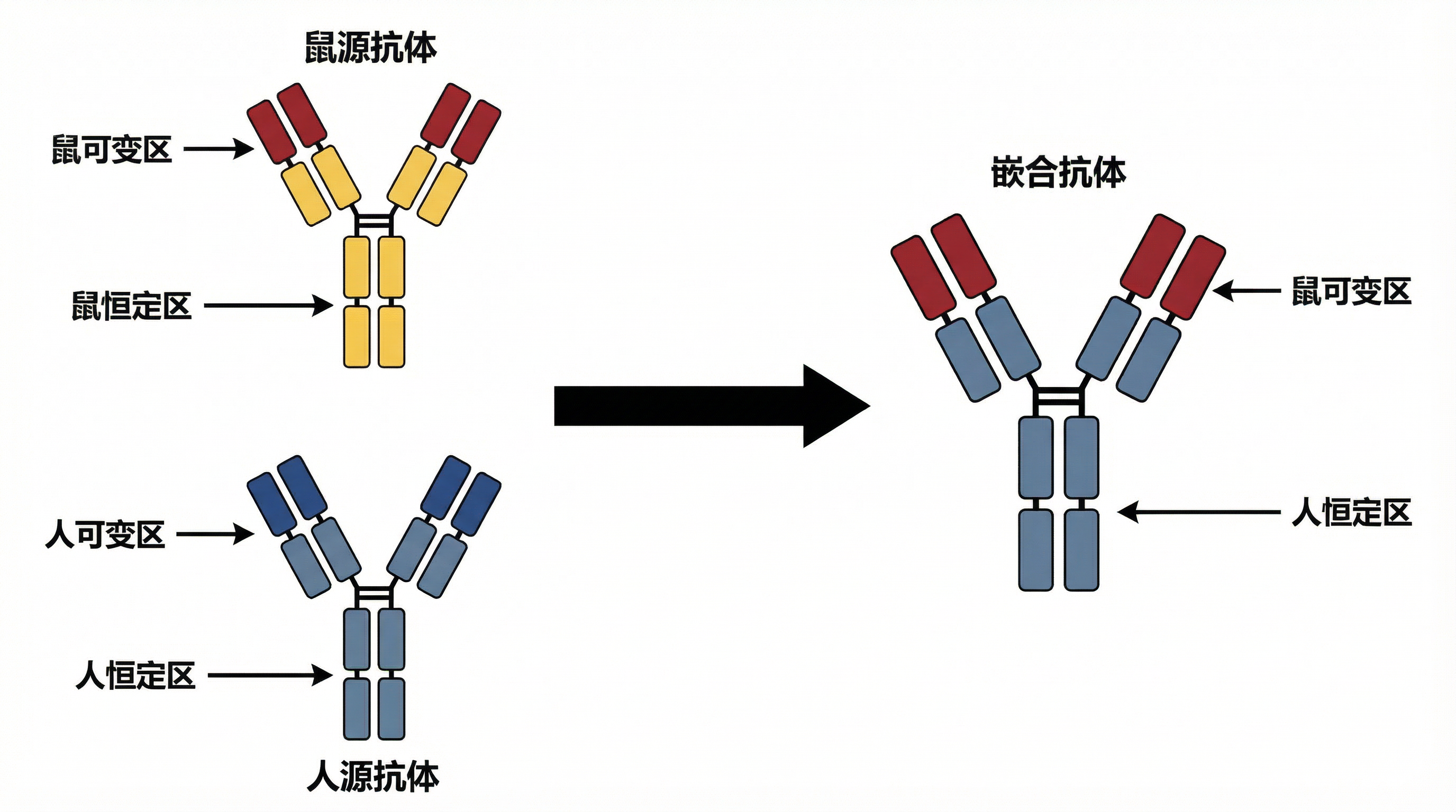
将这张抗体嵌合改造示意图调整为更严谨规范的学术期刊插图风格,线条清晰规整,配色专业简洁,标注准确,整体符合学术论文图表的严谨规范要求,保留原有结构逻辑和中文标注
GD401800062
2026.04.03
做同款
保留原图中的橙紫色抗体Y型结构元素,将原图片深紫色星空背景替换为干净柔和的浅淡浅色背景,保持元素不变
Blue_u
去除现有浅蓝色背景改为纯白色背景,提高整体颜色饱和度,让蓝色和粉色更加鲜艳,补全图像边缘缺失的抗体分子部分,保持抗体分子3D立体果冻质感,整体完整清晰
Blue_u
CAR-T细胞治疗示意图,展示改造后的免疫T细胞攻击癌细胞过程,现代医疗生物科技风格
6anhcooi9YTJZOg-HB
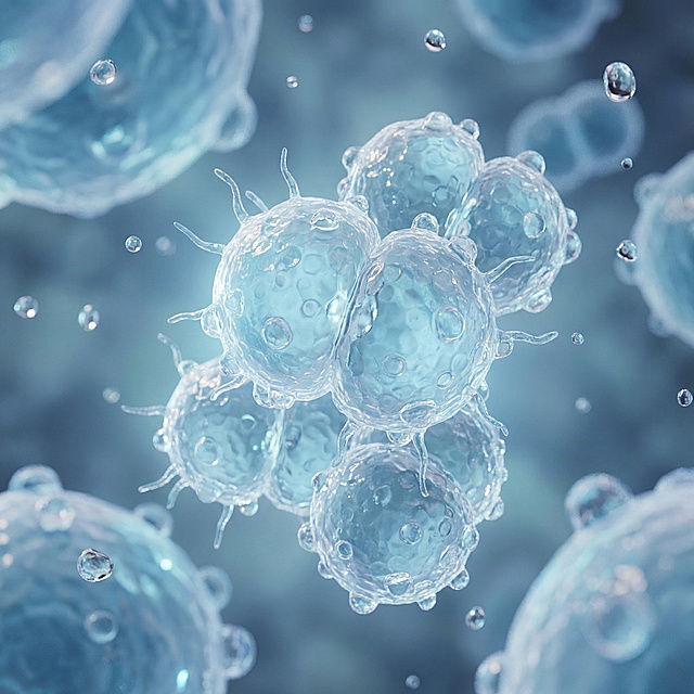
参考原图风格,将主体替换为脑膜炎球菌,保持透明质感的3D生物结构,浅蓝清新色调,微观显微镜视角,高清医疗可视化渲染,水珠光影效果,景深效果
caimin
删掉
1qunkq4t2gsNLOlA-HB
四周的黑色,加上些淡蓝的点点光芒
123lff
一颗巨大的半透明紫色球菌剪影,球体内部不是细胞结构,而是一间暖光照射的ICU病房场景剪影——病床、监护仪波形、一位医生俯身的轮廓;菌体下方一只戴蓝色手套的手轻轻捧着这颗菌体,如同捧着易碎的珍贵之物;深蓝渐变背景,只有菌体内部透出暖金色光;极简高级感,象征主义风格,干净无多余元素,适合PPT素养目标配图。
GD220794368
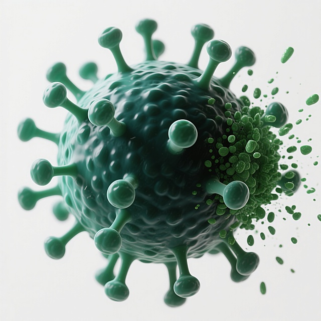
3D渲染的病毒微观结构,整体呈球形,表面布满凹凸不平的颗粒状纹理,主体为深绿色并带有浅绿色渐变,多个棒状突起物分布于球体表面,突起顶端呈圆形膨大,右侧有部分绿色颗粒状物质飞溅散开,高光与阴影对比强烈,光源来自左上方,呈现出高度写实的生物医学质感,白色背景,科学可视化风格,细节丰富,立体感强。
1qax0tk1gf1kvk3vu-HB
将深色背景修改为浅色背景,保留蓝绿色的病毒和DNA图形元素,整体保持自然协调,呈现高级清透的视觉效果
炸毛羊
参考图中的免疫学机制图风格(左侧胸腺Thymus区域、右侧外周Periphery区域、细胞卡通图标、箭头信号通路、分子标注、蛋白标记框),生成一张"免疫调节与Treg细胞分化机制模型"图。画面布局:左上为胸腺(Thymus)区,展示TEC细胞、初始T细胞分化为nTreg(天然调节性T细胞,Foxp3+红色细胞),标注CD25、Foxp3、CD39/73、LAG-3、CTLA-4、CD40、CD80/86、OX40、GITR等表面分子;右侧为外周(Periphery)区,展示iTreg(诱导性调节性T细胞)分化通路:包括Tconv(常规T细胞)在TGF-β、IL-10、IL-35作用下分化为Foxp3+ iTreg,Tr1细胞(分泌IL-10),Th3细胞(分泌TGF-β);下方展示APC抗原呈递细胞、DC树突状细胞(CD103+)与Treg的相互作用,标注GPR43/GPR109A、SCFA短链脂肪酸、视黄酸RA、CCR9/α4β7归巢受体、向GALT肠道相关淋巴组织迁移;右侧展示Treg的外周维持机制(IL-2、TGF-β、Foxp3+)。整体风格:细胞用彩色圆形卡通图标表示,分子用小圆点/三角形表示,黑色箭头指示分化和信号方向,白色方框标注分子标记,浅米色分区背景,绿色边框,专业学术示意图风格,英文标注为主,顶部大标题"免疫调节与Treg细胞分化机制模型"。
绎雪雪
Ultra-detailed scientific illustration, flat vector style, showcasing the process of "Triparental Mating" for bacterial plasmid transfer. The image is divided into three clear stages from left to right. Stage 1: Three distinct bacterial cells in close proximity. 1 Donor E. coli: Contains a "mobilizable plasmid" with a visible "gene of interest" (like a fluorescent protein or antibiotic resistance marker) and an "oriT" site. 2 Helper E. coli: Contains a "conjugative plasmid" with visible "tra genes" (transfer genes) and pilus structures. 3 Recipient bacterium (e.g., a Rhizobium cell): Has a different cell wall shape, labeled, with no plasmid inside. Stage 2: The helper E. coli extends a "pilus" (a thin tube) to attach to the donor E. coli. The conjugative plasmid's tra genes mobilize the mobilizable plasmid. Show a clear arrow indicating the transfer of the mobilizable plasmid FROM the donor TO the helper cell. Stage 3: The helper E. coli, now carrying the mobilizable plasmid, extends a new pilus to attach to the recipient bacterium. Show a second arrow indicating the final transfer of the mobilizable plasmid INTO the recipient. The recipient bacterium now contains the plasmid. Use a distinct, consistent color scheme: Donor is Blue, Helper is Orange, Recipient is Green, Mobilizable Plasmid is Red, Conjugative Plasmid is Yellow. The background is clean white. All key elements have clear labels with leader lines. The style is educational, schematic, and extremely clear for a presentation slide.
Poiuyt·
消除图片右下角的蓝色方块,保持整体科技生物医疗风格、浅蓝色调以及其他所有原内容不变,只移除右下角的蓝色方块区域,用和周围一致的背景填充
166****1440
一颗细菌的剖面示意图,左右两半完全对称但结构截然不同;左半为紫色厚壁,壁层致密如树轮,内部锁满深紫色颗粒状结晶紫-碘复合物;右半为红色薄壁,壁层疏松可见孔隙,紫色颗粒正从孔隙中向外流失滴落;中央竖直线分割左右,底部一行小字“结构决定结果”;极简矢量图标风,白底,线条干净清晰,适合PPT图标使用。
GD220794368
超写实风格医疗背景,主色调为科技蓝与深蓝渐变,柔和的侧顶光源营造出明亮通透的氛围,光线在玻璃器皿上形成清晰的高光与细腻阴影。前景中,戴着白色无菌手套的手正小心翼翼地握着一支装有透明液体的玻璃试管,手指关节自然弯曲,手套纹理清晰可见。背景中虚化的实验室环境里,多个不同形状的玻璃烧杯与试管整齐排列,后方叠加着动态的蓝色医疗数据波形图、DNA双螺旋结构和医疗图表的半透明全息投影效果,整体呈现出科技感与专业性并存的氛围。画面构图采用三分法,手部与试管位于视觉中心偏右位置,背景元素有序分布,形成层次感强的视觉引导。
1qax1093jb1ehaeqi-HB
亦是恩赐亦是劫か
将图中中间发光的橙红色神经元改成蓝色,保持整体画面结构和其他元素不变
朱朱biu特否
为瑞科生物公司设计一个高大上的蓝色圆形图标,主题是疫苗知识,图标内部不出现任何文字,以病毒盾牌简约符号表现,图标整体背景为纯白色,主色调为蓝色,设计风格简约高端大气,保持整体色调统一,线条流畅,生物科技感,健康医疗主题
1qfkdobz7hFNboRA-HB
参考图片风格,重新生成画面内容「图片1」帮我把文字全部抹除
故巷晚风
科学可视化3D示意图,展示核酸四面体DNA纳米结构,四个顶点的DNA折叠成立体框架,蓝色半透明分子结构,背景简洁,生物医药科技风格,高清
1r2dv8wr5nAVFdog-HB
国家级职业院校比赛专用PPT封面主视觉背景图,16:9尺寸,高清300DPI矢量精细画质,无水印无文字。主题:医学检验技术专业《细菌的革兰氏染色实验》信息化教学说课,主题标语「智辨阴阳,染求真知」。风格:医学专业学术感,前沿信息化科技感,赛事级高级仪式感,立体层次,光影质感。色彩体系:哑光高级灰白渐变基底,深海科技蓝做对称光影渐变,点缀结晶紫深紫(G+菌色)、番红砖红(G-菌色),低饱和高级质感,色彩过渡柔和,无高饱和刺眼色块。构图元素:对称庄重构图,上下边缘延伸沉浸式微观场景,左右两侧半透明3D立体G+紫色球菌和G-红色杆菌,柔焦显微镜油镜光影,虚拟仿真界面极简科技线条,AI数据流轻量线条,细菌细胞壁结构极简线稿,所有元素低透明度景深处理,画面正中心预留75%以上无干扰纯净渐变区域,不遮挡文字信息,无多余元素。细节质感:轻量径向光效,精致细线条分割,哑光磨砂渐变蒙版,拒绝平涂大白底,庄重大气,医学严谨+科技感,适配赛事投影,暗部不发黑亮部不曝光。只生成背景,不要有任何文字。
肥大大
简化图片,减少图片中的的元素,实现更加简洁的风格
yukkkkkki
顺着图片中的光线添加基因序列生物元素,保留原有的蓝色科技光效和波浪曲线结构,整体风格保持不变
白皙胜雪
3D渲染风格的生物医学微观背景,以紫色和蓝色为主色调,呈现出高度写实的细胞与分子结构。画面中心是一个半透明的蓝色细胞膜包裹着深紫色细胞核的细胞,悬浮在纹理粗糙、布满小孔和凸起的紫色细胞表面上。周围有多条螺旋状的DNA链,呈现出蓝紫色渐变,有的缠绕在细胞表面,有的向背景延伸。背景为渐变的浅蓝色,营造出深邃、神秘的科学氛围。整体光影柔和,细胞表面有细腻的高光和阴影,突出立体感和材质的颗粒感。构图采用近景特写,主体清晰,背景元素逐渐虚化,形成自然的景深效果,展现出微观世界的精密与奇妙。
1qax10ld9d16ttn69-HB
依照下面的话绘制一个流程图:1.细胞间相互作用的分子基础(细胞间接触和连接的研究):利用免疫荧光染色、共聚焦显微镜等技术,观察周细胞与内皮细胞、星形胶质细胞之间的接触和连接方式,包括紧密连接、桥粒连接、缝隙连接等。分析这些细胞间连接的组成成分和结构特点,探讨其在维持BBB功能中的作用。 2.细胞外环境因素对周细胞功能的调节(血管生成因子的影响):研究血管生成因子(如VEGF、bFGF)对周细胞功能的调节作用。通过添加不同浓度的血管生成因子,观察其对周细胞增殖、迁移、分化的影响,以及对BBB通透性和Aβ清除的间接作用。
1qnph8rssz83yZJA-HB
帮我生成50张关于月亮或者星球的图片
柠檬真甜