
创意预览图

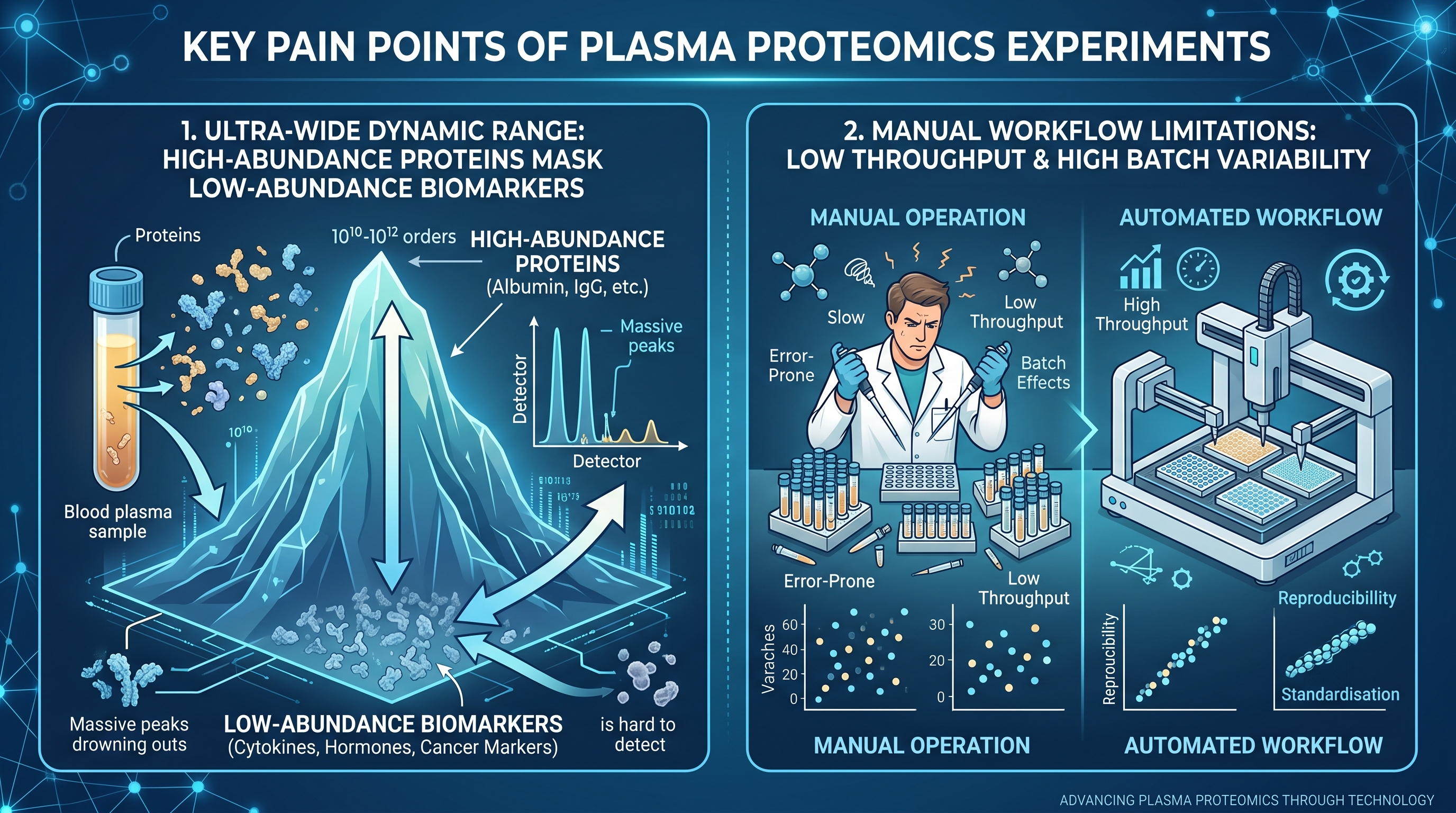
横版信息图,展示血浆蛋白组实验两大核心痛点:1.蛋白动态范围跨度极大,低丰度标志物难以检出,图示高丰度蛋白掩盖低丰度蛋白;2.手工处理通量低、批次差异严重,对比手工操作和自动化流程差异,科技风,蓝色商

用户头像

1q0vx9wwh3s...

2026.06.24
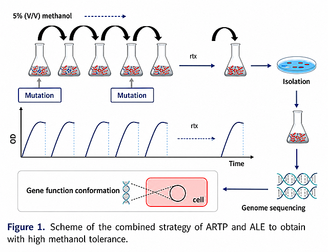
Ultra-detailed scientific illustration, flat vector style, showcasing the process of "Triparental Mating" for bacterial plasmid transfer. The image is divided into three clear stages from left to right. Stage 1: Three distinct bacterial cells in close proximity. 1 Donor E. coli: Contains a "mobilizable plasmid" with a visible "gene of interest" (like a fluorescent protein or antibiotic resistance marker) and an "oriT" site. 2 Helper E. coli: Contains a "conjugative plasmid" with visible "tra genes" (transfer genes) and pilus structures. 3 Recipient bacterium (e.g., a Rhizobium cell): Has a different cell wall shape, labeled, with no plasmid inside. Stage 2: The helper E. coli extends a "pilus" (a thin tube) to attach to the donor E. coli. The conjugative plasmid's tra genes mobilize the mobilizable plasmid. Show a clear arrow indicating the transfer of the mobilizable plasmid FROM the donor TO the helper cell. Stage 3: The helper E. coli, now carrying the mobilizable plasmid, extends a new pilus to attach to the recipient bacterium. Show a second arrow indicating the final transfer of the mobilizable plasmid INTO the recipient. The recipient bacterium now contains the plasmid. Use a distinct, consistent color scheme: Donor is Blue, Helper is Orange, Recipient is Green, Mobilizable Plasmid is Red, Conjugative Plasmid is Yellow. The background is clean white. All key elements have clear labels with leader lines. The style is educational, schematic, and extremely clear for a presentation slide.Ultra-detailed scientific illustration, flat vector style, showcasing the process of "Triparental Mating" for bacterial plasmid transfer. The image is divided into three clear stages from left to right. Stage 1: Three distinct bacterial cells in close proximity. 1 Donor E. coli: Contains a "mobilizable plasmid" with a visible "gene of interest" (like a fluorescent protein or antibiotic resistance marker) and an "oriT" site. 2 Helper E. coli: Contains a "conjugative plasmid" with visible "tra genes" (transfer genes) and pilus structures. 3 Recipient bacterium (e.g., a Rhizobium cell): Has a different cell wall shape, labeled, with no plasmid inside. Stage 2: The helper E. coli extends a "pilus" (a thin tube) to attach to the donor E. coli. The conjugative plasmid's tra genes mobilize the mobilizable plasmid. Show a clear arrow indicating the transfer of the mobilizable plasmid FROM the donor TO the helper cell. Stage 3: The helper E. coli, now carrying the mobilizable plasmid, extends a new pilus to attach to the recipient bacterium. Show a second arrow indicating the final transfer of the mobilizable plasmid INTO the recipient. The recipient bacterium now contains the plasmid. Use a distinct, consistent color scheme: Donor is Blue, Helper is Orange, Recipient is Green, Mobilizable Plasmid is Red, Conjugative Plasmid is Yellow. The background is clean white. All key elements have clear labels with leader lines. The style is educational, schematic, and extremely clear for a presentation slide.
生成一张国际会议学术海报,主题关于外泌体作为天然化合物纳米载体的研究,遵循参考图片风格,蓝色系专业生物科技风格,三列式布局。 标题为 "Exosomes as Advanced Nanocarriers for Natural Compounds: From Biogenesis to Therapeutic Applications" 作者:Dan Yu,联系方式:×××××× 左列:1. Introduction:定义外泌体是30-150nm内体来源囊泡,细胞间通讯关键分子,临床理想标志物,总结外泌体生物发生和作为天然化合物递送载体潜力;左列下半部分:2. Exosome Biogenesis:源自晚期内体→多泡体,包含ESCRT依赖和ESCRT非依赖途径,多泡体与质膜融合释放,由Rab GTPases等调控,在此模块添加外泌体形成机制流程示意图 中列上:3. Exosome Composition:表面富含四跨膜蛋白CD9、CD63、CD81等,管腔包含蛋白质HSP70、HSP90、酶、RNA等,能量依赖内吞摄取;中列下:4. Why Exosomes? 优势:生物相容性好低毒性、免疫逃逸、靶向能力、多功能性可递送多种 payload 右列上:5. Delivery of Natural Compounds案例展示:姜黄素抗炎免疫调节内皮保护,EGCG诱导巨噬细胞表型转变抑制肿瘤,在此模块放置靶向设计或姜黄素作用机制示意图;右列下:6. Challenges & Future Perspectives:纯化标准化挑战、生产 scalability、装载效率、药代动力学稳定性,展望外泌体作为下一代给药系统 bridging传统医学和精准治疗。 整体英文表述,专业清晰,学术风格,蓝色配色,布局三列式从左到右背景/机制→成分/优势→药物递送案例/结论。生成一张国际会议学术海报,主题关于外泌体作为天然化合物纳米载体的研究,遵循参考图片风格,蓝色系专业生物科技风格,三列式布局。
标题为 "Exosomes as Advanced Nanocarriers for Natural Compounds: From Biogenesis to Therapeutic Applications"
作者:Dan Yu,联系方式:××××××
左列:1. Introduction:定义外泌体是30-150nm内体来源囊泡,细胞间通讯关键分子,临床理想标志物,总结外泌体生物发生和作为天然化合物递送载体潜力;左列下半部分:2. Exosome Biogenesis:源自晚期内体→多泡体,包含ESCRT依赖和ESCRT非依赖途径,多泡体与质膜融合释放,由Rab GTPases等调控,在此模块添加外泌体形成机制流程示意图
中列上:3. Exosome Composition:表面富含四跨膜蛋白CD9、CD63、CD81等,管腔包含蛋白质HSP70、HSP90、酶、RNA等,能量依赖内吞摄取;中列下:4. Why Exosomes? 优势:生物相容性好低毒性、免疫逃逸、靶向能力、多功能性可递送多种 payload
右列上:5. Delivery of Natural Compounds案例展示:姜黄素抗炎免疫调节内皮保护,EGCG诱导巨噬细胞表型转变抑制肿瘤,在此模块放置靶向设计或姜黄素作用机制示意图;右列下:6. Challenges & Future Perspectives:纯化标准化挑战、生产 scalability、装载效率、药代动力学稳定性,展望外泌体作为下一代给药系统 bridging传统医学和精准治疗。
整体英文表述,专业清晰,学术风格,蓝色配色,布局三列式从左到右背景/机制→成分/优势→药物递送案例/结论。