
显微镜下的脓细胞,医学插画风格,清晰展示细胞结构,中性粒细胞吞… - 图片创意预览

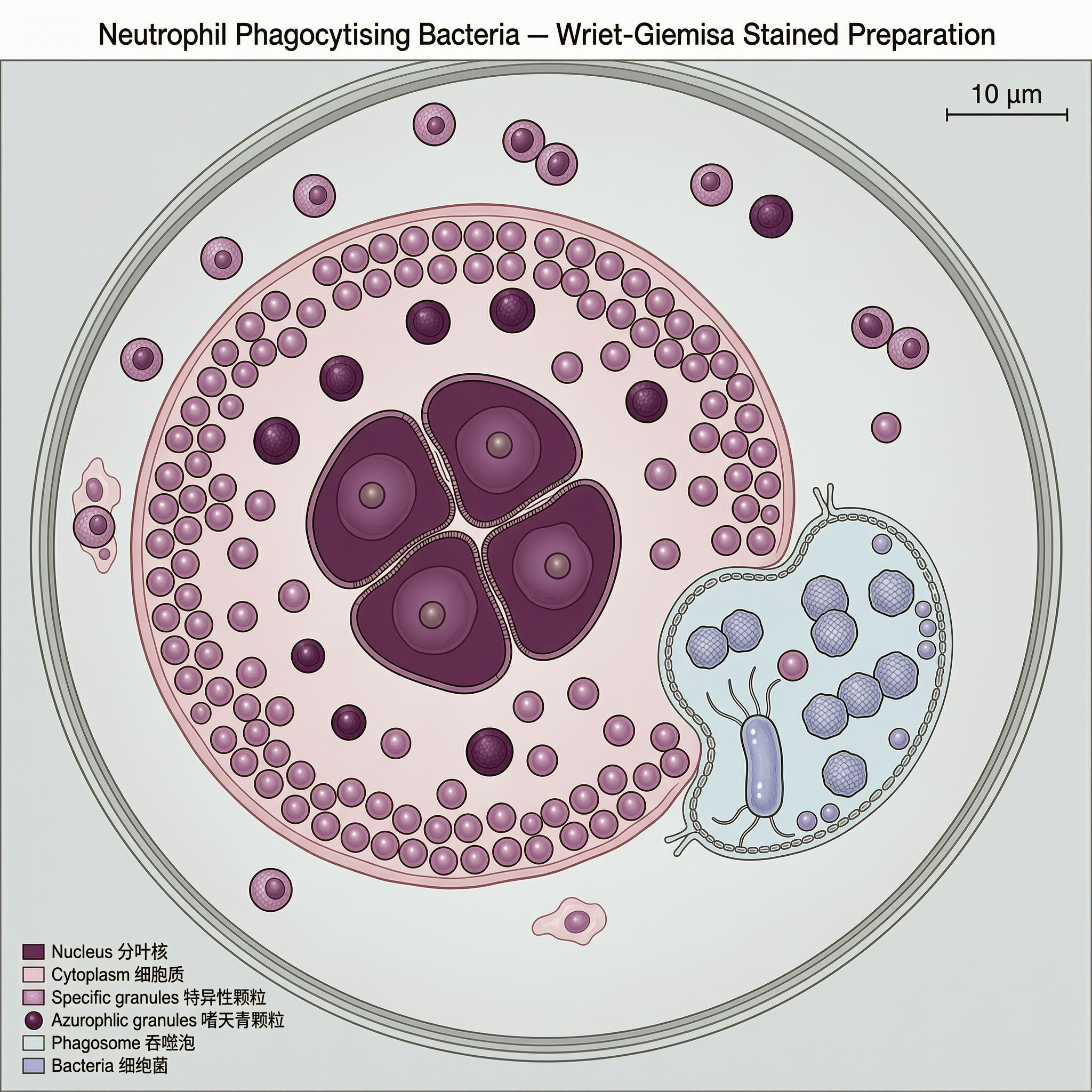
显微镜下的脓细胞,医学插画风格,清晰展示细胞结构,中性粒细胞吞…

显微镜下的脓细胞,医学插画风格,清晰展示细胞结构,中性粒细胞吞噬细菌的形态,细胞质内可见颗粒,分叶核明显,周围有细菌和组织碎片,科学准确的生物医学插图,高分辨率,细节丰富,专业医学绘图风格

用户头像

南絮58657

2026.09.19
Ultra-detailed scientific illustration, flat vector style, showcasing the process of "Triparental Mating" for bacterial plasmid transfer. The image is divided into three clear stages from left to right. Stage 1: Three distinct bacterial cells in close proximity. 1 Donor E. coli: Contains a "mobilizable plasmid" with a visible "gene of interest" (like a fluorescent protein or antibiotic resistance marker) and an "oriT" site. 2 Helper E. coli: Contains a "conjugative plasmid" with visible "tra genes" (transfer genes) and pilus structures. 3 Recipient bacterium (e.g., a Rhizobium cell): Has a different cell wall shape, labeled, with no plasmid inside. Stage 2: The helper E. coli extends a "pilus" (a thin tube) to attach to the donor E. coli. The conjugative plasmid's tra genes mobilize the mobilizable plasmid. Show a clear arrow indicating the transfer of the mobilizable plasmid FROM the donor TO the helper cell. Stage 3: The helper E. coli, now carrying the mobilizable plasmid, extends a new pilus to attach to the recipient bacterium. Show a second arrow indicating the final transfer of the mobilizable plasmid INTO the recipient. The recipient bacterium now contains the plasmid. Use a distinct, consistent color scheme: Donor is Blue, Helper is Orange, Recipient is Green, Mobilizable Plasmid is Red, Conjugative Plasmid is Yellow. The background is clean white. All key elements have clear labels with leader lines. The style is educational, schematic, and extremely clear for a presentation slide.Ultra-detailed scientific illustration, flat vector style, showcasing the process of "Triparental Mating" for bacterial plasmid transfer. The image is divided into three clear stages from left to right. Stage 1: Three distinct bacterial cells in close proximity. 1 Donor E. coli: Contains a "mobilizable plasmid" with a visible "gene of interest" (like a fluorescent protein or antibiotic resistance marker) and an "oriT" site. 2 Helper E. coli: Contains a "conjugative plasmid" with visible "tra genes" (transfer genes) and pilus structures. 3 Recipient bacterium (e.g., a Rhizobium cell): Has a different cell wall shape, labeled, with no plasmid inside. Stage 2: The helper E. coli extends a "pilus" (a thin tube) to attach to the donor E. coli. The conjugative plasmid's tra genes mobilize the mobilizable plasmid. Show a clear arrow indicating the transfer of the mobilizable plasmid FROM the donor TO the helper cell. Stage 3: The helper E. coli, now carrying the mobilizable plasmid, extends a new pilus to attach to the recipient bacterium. Show a second arrow indicating the final transfer of the mobilizable plasmid INTO the recipient. The recipient bacterium now contains the plasmid. Use a distinct, consistent color scheme: Donor is Blue, Helper is Orange, Recipient is Green, Mobilizable Plasmid is Red, Conjugative Plasmid is Yellow. The background is clean white. All key elements have clear labels with leader lines. The style is educational, schematic, and extremely clear for a presentation slide.