
创意预览图

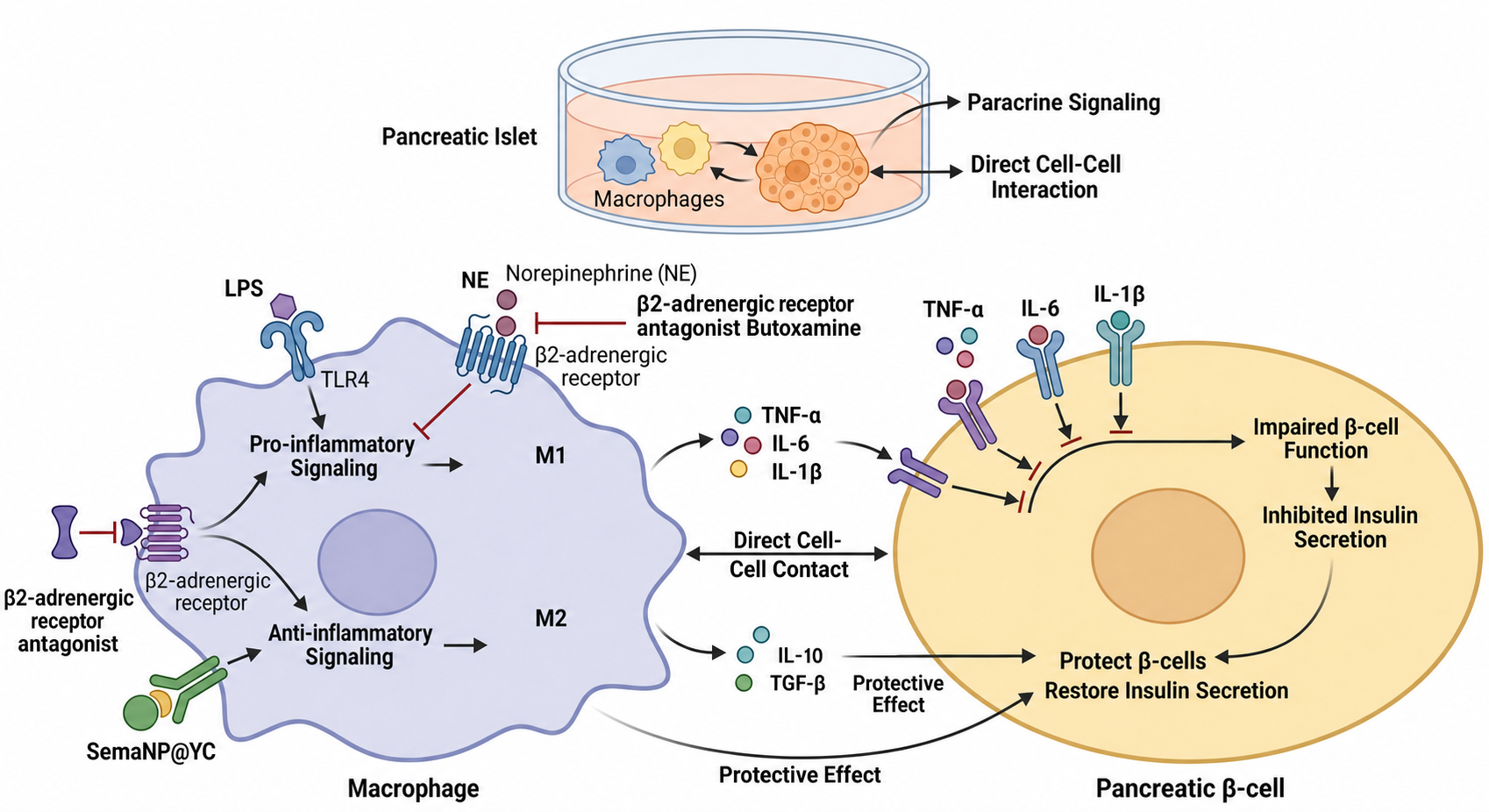
删除最上方标题文字,删除(e.g.,NF-kB)、(e.g.,cAMP)、Pro-inflammatory Polarization、Anti-inflammatory Polarization文字,

Ultra-detailed scientific illustration, flat vector style, showcasing the process of "Triparental Mating" for bacterial plasmid transfer. The image is divided into three clear stages from left to right. Stage 1: Three distinct bacterial cells in close proximity. 1 Donor E. coli: Contains a "mobilizable plasmid" with a visible "gene of interest" (like a fluorescent protein or antibiotic resistance marker) and an "oriT" site. 2 Helper E. coli: Contains a "conjugative plasmid" with visible "tra genes" (transfer genes) and pilus structures. 3 Recipient bacterium (e.g., a Rhizobium cell): Has a different cell wall shape, labeled, with no plasmid inside. Stage 2: The helper E. coli extends a "pilus" (a thin tube) to attach to the donor E. coli. The conjugative plasmid's tra genes mobilize the mobilizable plasmid. Show a clear arrow indicating the transfer of the mobilizable plasmid FROM the donor TO the helper cell. Stage 3: The helper E. coli, now carrying the mobilizable plasmid, extends a new pilus to attach to the recipient bacterium. Show a second arrow indicating the final transfer of the mobilizable plasmid INTO the recipient. The recipient bacterium now contains the plasmid. Use a distinct, consistent color scheme: Donor is Blue, Helper is Orange, Recipient is Green, Mobilizable Plasmid is Red, Conjugative Plasmid is Yellow. The background is clean white. All key elements have clear labels with leader lines. The style is educational, schematic, and extremely clear for a presentation slide.Ultra-detailed scientific illustration, flat vector style, showcasing the process of "Triparental Mating" for bacterial plasmid transfer. The image is divided into three clear stages from left to right. Stage 1: Three distinct bacterial cells in close proximity. 1 Donor E. coli: Contains a "mobilizable plasmid" with a visible "gene of interest" (like a fluorescent protein or antibiotic resistance marker) and an "oriT" site. 2 Helper E. coli: Contains a "conjugative plasmid" with visible "tra genes" (transfer genes) and pilus structures. 3 Recipient bacterium (e.g., a Rhizobium cell): Has a different cell wall shape, labeled, with no plasmid inside. Stage 2: The helper E. coli extends a "pilus" (a thin tube) to attach to the donor E. coli. The conjugative plasmid's tra genes mobilize the mobilizable plasmid. Show a clear arrow indicating the transfer of the mobilizable plasmid FROM the donor TO the helper cell. Stage 3: The helper E. coli, now carrying the mobilizable plasmid, extends a new pilus to attach to the recipient bacterium. Show a second arrow indicating the final transfer of the mobilizable plasmid INTO the recipient. The recipient bacterium now contains the plasmid. Use a distinct, consistent color scheme: Donor is Blue, Helper is Orange, Recipient is Green, Mobilizable Plasmid is Red, Conjugative Plasmid is Yellow. The background is clean white. All key elements have clear labels with leader lines. The style is educational, schematic, and extremely clear for a presentation slide.
16:9横版植物细胞通路图,分细胞外、细胞质、细胞核、叶绿体、线粒体、液泡区域。细胞质中心是亮金橙色SnRK1复合物,是核心调控器。绿色实心箭头为激活通路,红色钝线为抑制通路,黄色带“P”小圆圈是磷酸化,紫色带“Ub”小圆圈是泛素化。顶部中心:乌云降雨、水位图标代表环境触发,文字“Energy Stress / Carbon Starvation / Low Photosynthesis”,粗箭头指向SnRK1,使其从灰色 inactive 变为发光激活态。左侧细胞质:绘制HMGR酶,SnRK1连红色钝线带“P”(Ser577)指向它,灰色划掉的通路“Acetyl-CoA → MVA Pathway → Sterols/Brassinosteroids”;绘制PAL酶,SnRK1触发紫色“Ub”连接到PAL,PAL指向垃圾桶状蛋白酶体,灰色划掉通路“Phenylalanine → Lignin”,文字“Reduces Carbon Consumption”。右上方细胞质和叶绿体:细胞质中SnRK1给F2KP加“P”(Ser306),14-3-3蛋白结合磷酸化的F2KP(红色钝线抑制),F2KP失活使F-2,6-BP减少,减少的F-2,6-BP解除对cyFBPase的抑制(红色虚线划掉),cyFBPase将F-1,6-BP转化为F-6-P;SnRK1直接磷酸化抑制蔗糖合成酶。绿色叶绿体中,淀粉颗粒在BAM1/3、BAM9酶作用下降解为麦芽糖,细胞核箭头指向叶绿体中的BAM9。细胞核区域:SnRK1催化亚基进入细胞核,激活DNA链上的bZIP TFs和C/S1-bZIPs。基因表达:SUC1表达被抑制(红叉),质膜上SUC1转运体变灰;BAM9上调(箭头指向叶绿体);DIN6 (ASN1)上调转化为天冬酰胺(Asn);CAT6上调,质膜上CAT6转运体将天冬酰胺/氨基酸泵出细胞。底部区域:细胞质/液泡中,SnRK1(KIN10亚基)给ATG6加“P”启动自噬体(新月形膜),给FREE1加“P”促进自噬体闭合,自噬体与液泡融合释放氨基酸(AAs);线粒体释放FLZ(SnRK1抑制剂),FLZ被选择性自噬吞噬在液泡降解。右下角线粒体:亮氨酸、异亮氨酸、缬氨酸进入线粒体,代谢链BCAT2 → BCKDH → IVDH → MCCA/B生成乙酰辅酶A进入三羧酸循环,IVDH连侧箭头显示电子(e-)到ETF → ETFQO → 泛醌(UQ)进入电子传递链;细胞核(C/S1-bZIPs)箭头上调ETFQO、BCAT2、BCKDH等转录。用半透明彩色背景框分组:蓝色框“Carbon Flow Restriction”,绿色框“Alternative Energy (BCAAs & Autophagy)”,橙色框“Carbohydrate Homeostasis”。SnRK1最大最突出,主代谢流用粗箭头,调控信号用细线,文字简洁,酶用酶形状标注。16:9横版植物细胞通路图,分细胞外、细胞质、细胞核、叶绿体、线粒体、液泡区域。细胞质中心是亮金橙色SnRK1复合物,是核心调控器。绿色实心箭头为激活通路,红色钝线为抑制通路,黄色带“P”小圆圈是磷酸化,紫色带“Ub”小圆圈是泛素化。顶部中心:乌云降雨、水位图标代表环境触发,文字“Energy Stress / Carbon Starvation / Low Photosynthesis”,粗箭头指向SnRK1,使其从灰色 inactive 变为发光激活态。左侧细胞质:绘制HMGR酶,SnRK1连红色钝线带“P”(Ser577)指向它,灰色划掉的通路“Acetyl-CoA → MVA Pathway → Sterols/Brassinosteroids”;绘制PAL酶,SnRK1触发紫色“Ub”连接到PAL,PAL指向垃圾桶状蛋白酶体,灰色划掉通路“Phenylalanine → Lignin”,文字“Reduces Carbon Consumption”。右上方细胞质和叶绿体:细胞质中SnRK1给F2KP加“P”(Ser306),14-3-3蛋白结合磷酸化的F2KP(红色钝线抑制),F2KP失活使F-2,6-BP减少,减少的F-2,6-BP解除对cyFBPase的抑制(红色虚线划掉),cyFBPase将F-1,6-BP转化为F-6-P;SnRK1直接磷酸化抑制蔗糖合成酶。绿色叶绿体中,淀粉颗粒在BAM1/3、BAM9酶作用下降解为麦芽糖,细胞核箭头指向叶绿体中的BAM9。细胞核区域:SnRK1催化亚基进入细胞核,激活DNA链上的bZIP TFs和C/S1-bZIPs。基因表达:SUC1表达被抑制(红叉),质膜上SUC1转运体变灰;BAM9上调(箭头指向叶绿体);DIN6 (ASN1)上调转化为天冬酰胺(Asn);CAT6上调,质膜上CAT6转运体将天冬酰胺/氨基酸泵出细胞。底部区域:细胞质/液泡中,SnRK1(KIN10亚基)给ATG6加“P”启动自噬体(新月形膜),给FREE1加“P”促进自噬体闭合,自噬体与液泡融合释放氨基酸(AAs);线粒体释放FLZ(SnRK1抑制剂),FLZ被选择性自噬吞噬在液泡降解。右下角线粒体:亮氨酸、异亮氨酸、缬氨酸进入线粒体,代谢链BCAT2 → BCKDH → IVDH → MCCA/B生成乙酰辅酶A进入三羧酸循环,IVDH连侧箭头显示电子(e-)到ETF → ETFQO → 泛醌(UQ)进入电子传递链;细胞核(C/S1-bZIPs)箭头上调ETFQO、BCAT2、BCKDH等转录。用半透明彩色背景框分组:蓝色框“Carbon Flow Restriction”,绿色框“Alternative Energy (BCAAs & Autophagy)”,橙色框“Carbohydrate Homeostasis”。SnRK1最大最突出,主代谢流用粗箭头,调控信号用细线,文字简洁,酶用酶形状标注。