Biomedical schematic of three c… - 图片创意预览

Biomedical schematic of three c…

Biomedical schematic of three core pathological mechanisms in infected wounds, in Nature Reviews sty

用户头像

GD442223348

2026.07.15
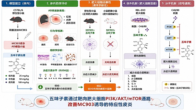
绘制一张符合《自然》期刊风格的高质量分子机制示意图,不是流程图,围绕五味子素缓解特应性皮炎的核心机制,字体大而清晰。 画面布局分为三大部分,从左到右逻辑递进: 左侧:MC903诱导的AD皮肤炎症场景。展示皮肤横截面,表皮增生、真皮层肥大细胞聚集活化,释放炎症介质(组胺、类胰蛋白酶、LTB4、IL-4、IL-13、TSLP、IgE),导致皮肤红斑、肿胀、瘙痒。标注"MC903-induced AD-like skin inflammation"。 中间:五味子素作用核心。展示五味子素(Schisandrin)的正确化学结构式,箭头指向肥大细胞,标注其靶向作用。肥大细胞内部绘制PI3K/AKT/mTOR信号通路:C48/80刺激激活PI3K磷酸化(Ser317)→AKT磷酸化(Ser473)→mTOR磷酸化(Ser2448)→肥大细胞活化脱颗粒。五味子素以抑制符号(T型线)作用于p-PI3K、p-AKT、p-mTOR三个磷酸化节点,总蛋白水平不变。标注总蛋白无显著变化。 右侧:治疗效果。展示治疗后皮肤恢复正常,肥大细胞减少,炎症介质水平下降,AD样皮损缓解。下方展示肥大细胞依赖性验证:野生型小鼠+五味子素→治疗效果显著;c-Kit肥大细胞缺失小鼠+五味子素→治疗效果减弱,证明作用依赖肥大细胞。 底部大标题:"Schisandrin alleviates atopic dermatitis by inhibiting mast cell activation via the PI3K/AKT/mTOR pathway" 整体风格:Nature期刊风格,配色高级协调,插图精美专业,字体大而清晰,线条流畅,有图例说明,适合顶刊发表。绘制一张符合《自然》期刊风格的高质量分子机制示意图,不是流程图,围绕五味子素缓解特应性皮炎的核心机制,字体大而清晰。

画面布局分为三大部分,从左到右逻辑递进:

左侧:MC903诱导的AD皮肤炎症场景。展示皮肤横截面,表皮增生、真皮层肥大细胞聚集活化,释放炎症介质(组胺、类胰蛋白酶、LTB4、IL-4、IL-13、TSLP、IgE),导致皮肤红斑、肿胀、瘙痒。标注"MC903-induced AD-like skin inflammation"。

中间:五味子素作用核心。展示五味子素(Schisandrin)的正确化学结构式,箭头指向肥大细胞,标注其靶向作用。肥大细胞内部绘制PI3K/AKT/mTOR信号通路:C48/80刺激激活PI3K磷酸化(Ser317)→AKT磷酸化(Ser473)→mTOR磷酸化(Ser2448)→肥大细胞活化脱颗粒。五味子素以抑制符号(T型线)作用于p-PI3K、p-AKT、p-mTOR三个磷酸化节点,总蛋白水平不变。标注总蛋白无显著变化。

右侧:治疗效果。展示治疗后皮肤恢复正常,肥大细胞减少,炎症介质水平下降,AD样皮损缓解。下方展示肥大细胞依赖性验证:野生型小鼠+五味子素→治疗效果显著;c-Kit肥大细胞缺失小鼠+五味子素→治疗效果减弱,证明作用依赖肥大细胞。

底部大标题:"Schisandrin alleviates atopic dermatitis by inhibiting mast cell activation via the PI3K/AKT/mTOR pathway"

整体风格:Nature期刊风格,配色高级协调,插图精美专业,字体大而清晰,线条流畅,有图例说明,适合顶刊发表。
Ultra-detailed scientific illustration, flat vector style, showcasing the process of "Triparental Mating" for bacterial plasmid transfer. The image is divided into three clear stages from left to right. Stage 1: Three distinct bacterial cells in close proximity. 1 Donor E. coli: Contains a "mobilizable plasmid" with a visible "gene of interest" (like a fluorescent protein or antibiotic resistance marker) and an "oriT" site. 2 Helper E. coli: Contains a "conjugative plasmid" with visible "tra genes" (transfer genes) and pilus structures. 3 Recipient bacterium (e.g., a Rhizobium cell): Has a different cell wall shape, labeled, with no plasmid inside. Stage 2: The helper E. coli extends a "pilus" (a thin tube) to attach to the donor E. coli. The conjugative plasmid's tra genes mobilize the mobilizable plasmid. Show a clear arrow indicating the transfer of the mobilizable plasmid FROM the donor TO the helper cell. Stage 3: The helper E. coli, now carrying the mobilizable plasmid, extends a new pilus to attach to the recipient bacterium. Show a second arrow indicating the final transfer of the mobilizable plasmid INTO the recipient. The recipient bacterium now contains the plasmid. Use a distinct, consistent color scheme: Donor is Blue, Helper is Orange, Recipient is Green, Mobilizable Plasmid is Red, Conjugative Plasmid is Yellow. The background is clean white. All key elements have clear labels with leader lines. The style is educational, schematic, and extremely clear for a presentation slide.Ultra-detailed scientific illustration, flat vector style, showcasing the process of "Triparental Mating" for bacterial plasmid transfer. The image is divided into three clear stages from left to right. Stage 1: Three distinct bacterial cells in close proximity. 1 Donor E. coli: Contains a "mobilizable plasmid" with a visible "gene of interest" (like a fluorescent protein or antibiotic resistance marker) and an "oriT" site. 2 Helper E. coli: Contains a "conjugative plasmid" with visible "tra genes" (transfer genes) and pilus structures. 3 Recipient bacterium (e.g., a Rhizobium cell): Has a different cell wall shape, labeled, with no plasmid inside. Stage 2: The helper E. coli extends a "pilus" (a thin tube) to attach to the donor E. coli. The conjugative plasmid's tra genes mobilize the mobilizable plasmid. Show a clear arrow indicating the transfer of the mobilizable plasmid FROM the donor TO the helper cell. Stage 3: The helper E. coli, now carrying the mobilizable plasmid, extends a new pilus to attach to the recipient bacterium. Show a second arrow indicating the final transfer of the mobilizable plasmid INTO the recipient. The recipient bacterium now contains the plasmid. Use a distinct, consistent color scheme: Donor is Blue, Helper is Orange, Recipient is Green, Mobilizable Plasmid is Red, Conjugative Plasmid is Yellow. The background is clean white. All key elements have clear labels with leader lines. The style is educational, schematic, and extremely clear for a presentation slide.