创意预览图

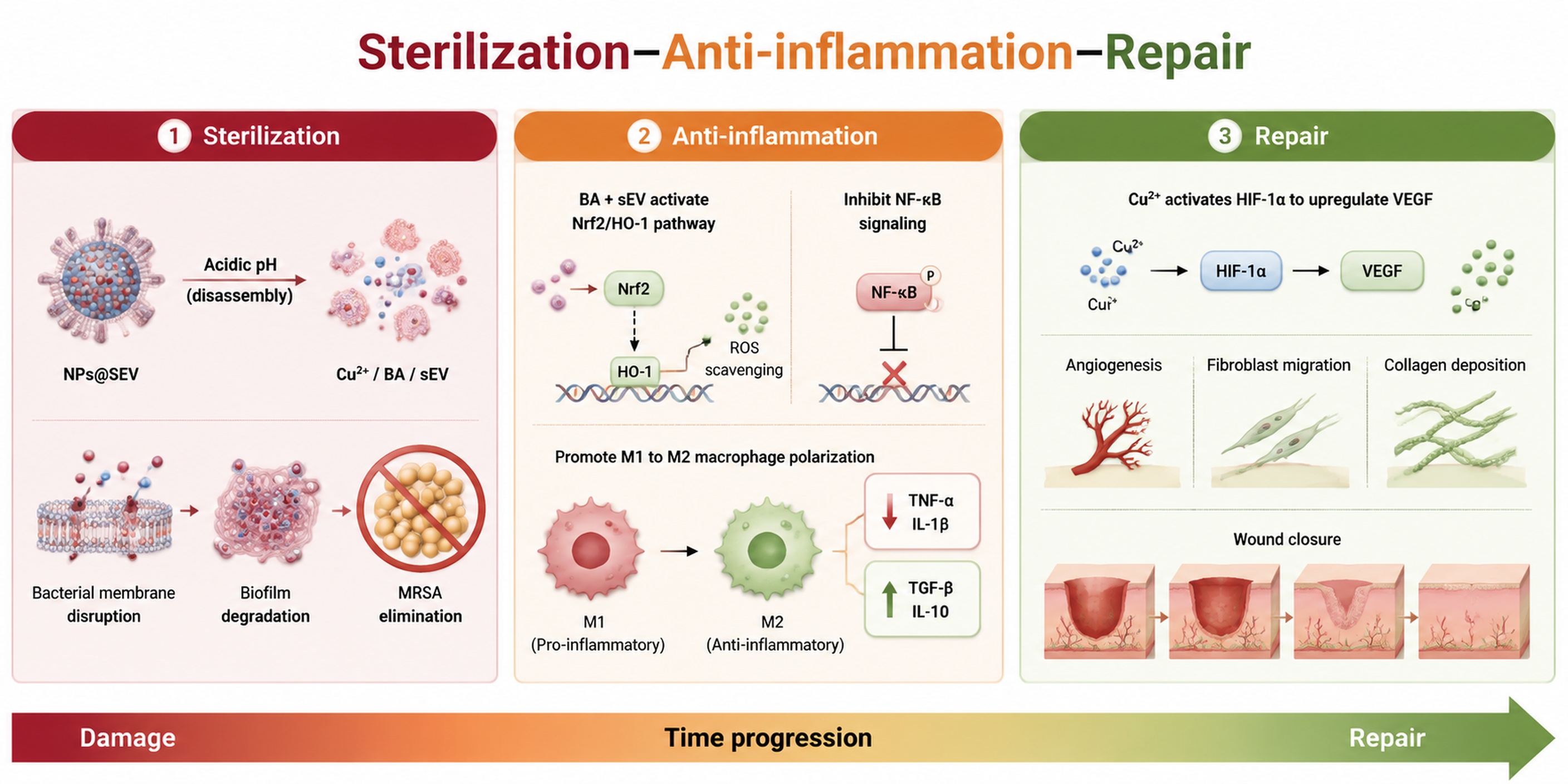
Tripartite synergistic therapeutic strategy diagram titled "sterilization-anti-inflammation-repair"

用户头像

GD442223348

2026.07.15
HPV相关宫颈病变的发生发展不仅受病毒癌蛋白E6/E7影响,还与宿主细胞表观遗传调控、炎症反应和程序性细胞死亡密切相关。持续性HPV感染可造成宫颈局部慢性炎症和免疫微环境改变,而这种长期炎症状态既可能促进异常细胞清除,也可能在持续激活后形成促病变进展的微环境。因此,除E6/E7-p53/pRb这一经典通路外,进一步解析表观转录调控与炎症性细胞死亡之间的关系,有助于拓展HPV相关宫颈病变机制研究的深度。 m6A修饰是真核mRNA中重要的表观转录修饰,可影响mRNA剪接、稳定性、翻译效率及降解过程。METTL3作为m6A甲基转移酶复合体中的核心“写入器”,在多种肿瘤中被证实可通过调控关键mRNA的m6A修饰影响细胞增殖、迁移、免疫反应和细胞死亡方式。宫颈癌相关研究也提示METTL3异常表达与肿瘤进展、侵袭转移和治疗反应存在关联,但其在HPV阳性宫颈细胞炎症性死亡调控中的具体作用仍未完全明确。 NLRP3炎症小体是先天免疫反应中的重要感受平台,可在细胞受到病原体相关分子、损伤信号或氧化应激刺激后被激活,并通过ASC招募和激活caspase-1,进一步促进IL-1β、IL-18成熟释放以及GSDMD切割,最终诱导焦亡。焦亡是一种具有炎症放大效应的程序性细胞死亡方式,在病毒感染、肿瘤免疫和慢性炎症中均具有重要作用。对于HPV阳性宫颈细胞而言,NLRP3炎症小体和焦亡既可能参与异常细胞清除,也可能通过持续炎症因子释放影响局部微环境。因此,阐明NLRP3炎症小体在HPV阳性宫颈细胞中的激活机制具有明确研究价值。 已有研究提示m6A修饰可参与NLRP3表达及炎症小体激活调控,但在HPV阳性宫颈细胞中,METTL3是否通过m6A修饰调控NLRP3 mRNA稳定性或翻译效率,并进一步影响caspase-1/GSDMD介导的焦亡,仍缺乏系统验证。本方向拟先利用GEO或TCGA等公共数据库分析METTL3、NLRP3及焦亡相关分子在宫颈病变和宫颈癌相关数据中的表达趋势,再通过METTL3敲低或抑制剂处理验证其对NLRP3、ASC、cleaved caspase-1、GSDMD-N、IL-1β、IL-18等指标的影响,并结合LDH释放、PI摄取和细胞死亡比例评估焦亡表型。进一步采用MeRIP-qPCR检测NLRP3 mRNA的m6A富集水平,并通过NLRP3过表达救援实验验证其在METTL3调控焦亡中的作用。该设计可形成“METTL3-m6A-NLRP3炎症小体-焦亡”的机制链条,为HPV阳性宫颈细胞炎症性死亡调控提供新的实验依据。 4. 研究假设 HPV持续感染相关宫颈细胞中,病毒癌蛋白和局部炎症刺激可能共同改变宿主细胞m6A表观转录修饰水平,使METTL3表达或活性升高。METTL3可能通过增强NLRP3 mRNA的m6A修饰,促进NLRP3 mRNA稳定性或翻译效率增加,进而激活NLRP3炎症小体,推动ASC募集、caspase-1活化、GSDMD切割以及IL-1β、IL-18成熟释放,最终影响HPV阳性宫颈细胞的焦亡水平和炎症放大反应。 本研究推测,METTL3-m6A-NLRP3轴是连接HPV相关表观转录异常与炎症性细胞死亡的重要分子通路。若METTL3敲低或抑制后,NLRP3 mRNA的m6A富集水平下降,NLRP3蛋白表达、cleaved caspase-1、GSDMD-N、IL-1β、IL-18、LDH释放和PI阳性细胞比例同步降低,则说明METTL3参与NLRP3炎症小体和焦亡调控。若NLRP3过表达能够部分恢复METTL3敲低后被抑制的焦亡表型,则可进一步证明NLRP3是METTL3调控HPV阳性宫颈细胞焦亡的关键下游节点。 5. 研究内容 5.1 生信分析METTL3、NLRP3及焦亡相关分子在宫颈病变中的表达特征 利用GEO或TCGA等公共数据库,分析METTL3、NLRP3、ASC、CASP1、GSDMD、IL1B、IL18等分子在正常宫颈、宫颈病变和宫颈癌相关数据中的表达趋势。通过差异表达分析、相关性分析和通路富集分析,判断METTL3、NLRP3及焦亡相关分子是否与HPV相关宫颈病变进展具有表达关联。若具备少量组织样本或病理蜡块条件,可采用免疫组化检测METTL3、NLRP3、GSDMD等指标作为补充验证,用于增强数据库分析结果的组织水平支持。 5.2 METTL3对NLRP3炎症小体和焦亡的调控作用 选取SiHa和HeLa细胞作为HPV阳性宫颈细胞模型,通过siRNA敲低METTL3或采用METTL3抑制剂处理,检测NLRP3、ASC、cleaved caspase-1、GSDMD-N、IL-1β、IL-18等炎症小体和焦亡相关指标变化。同步检测LDH释放、PI摄取和细胞死亡比例,判断METTL3降低后是否影响NLRP3炎症小体活化及焦亡水平。 5.3 METTL3通过m6A修饰调控NLRP3的机制验证 在METTL3敲低或过表达条件下,检测NLRP3 mRNA和蛋白表达变化。条件允许时,采用MeRIP-qPCR检测NLRP3 mRNA的m6A富集水平,观察METTL3是否影响NLRP3 mRNA上的m6A修饰。进一步通过NLRP3过表达救援实验,观察NLRP3是否能够部分恢复METTL3敲低后下降的焦亡相关指标,从而验证NLRP3在METTL3调控焦亡中的作用。 5.4 HPV E6/E7与METTL3-NLRP3轴的关联验证 在SiHa或HeLa细胞中沉默E6/E7,检测METTL3、NLRP3及焦亡相关指标变化;也可在C33A或正常宫颈上皮细胞中表达E6/E7,观察METTL3-NLRP3轴是否被激活。该部分用于连接HPV癌蛋白与METTL3-NLRP3焦亡轴之间的关系,增强研究的HPV疾病背景。 帮我根据这个研究方案生成一套完整的图片HPV相关宫颈病变的发生发展不仅受病毒癌蛋白E6/E7影响,还与宿主细胞表观遗传调控、炎症反应和程序性细胞死亡密切相关。持续性HPV感染可造成宫颈局部慢性炎症和免疫微环境改变,而这种长期炎症状态既可能促进异常细胞清除,也可能在持续激活后形成促病变进展的微环境。因此,除E6/E7-p53/pRb这一经典通路外,进一步解析表观转录调控与炎症性细胞死亡之间的关系,有助于拓展HPV相关宫颈病变机制研究的深度。
m6A修饰是真核mRNA中重要的表观转录修饰,可影响mRNA剪接、稳定性、翻译效率及降解过程。METTL3作为m6A甲基转移酶复合体中的核心“写入器”,在多种肿瘤中被证实可通过调控关键mRNA的m6A修饰影响细胞增殖、迁移、免疫反应和细胞死亡方式。宫颈癌相关研究也提示METTL3异常表达与肿瘤进展、侵袭转移和治疗反应存在关联,但其在HPV阳性宫颈细胞炎症性死亡调控中的具体作用仍未完全明确。
NLRP3炎症小体是先天免疫反应中的重要感受平台,可在细胞受到病原体相关分子、损伤信号或氧化应激刺激后被激活,并通过ASC招募和激活caspase-1,进一步促进IL-1β、IL-18成熟释放以及GSDMD切割,最终诱导焦亡。焦亡是一种具有炎症放大效应的程序性细胞死亡方式,在病毒感染、肿瘤免疫和慢性炎症中均具有重要作用。对于HPV阳性宫颈细胞而言,NLRP3炎症小体和焦亡既可能参与异常细胞清除,也可能通过持续炎症因子释放影响局部微环境。因此,阐明NLRP3炎症小体在HPV阳性宫颈细胞中的激活机制具有明确研究价值。
已有研究提示m6A修饰可参与NLRP3表达及炎症小体激活调控,但在HPV阳性宫颈细胞中,METTL3是否通过m6A修饰调控NLRP3 mRNA稳定性或翻译效率,并进一步影响caspase-1/GSDMD介导的焦亡,仍缺乏系统验证。本方向拟先利用GEO或TCGA等公共数据库分析METTL3、NLRP3及焦亡相关分子在宫颈病变和宫颈癌相关数据中的表达趋势,再通过METTL3敲低或抑制剂处理验证其对NLRP3、ASC、cleaved caspase-1、GSDMD-N、IL-1β、IL-18等指标的影响,并结合LDH释放、PI摄取和细胞死亡比例评估焦亡表型。进一步采用MeRIP-qPCR检测NLRP3 mRNA的m6A富集水平,并通过NLRP3过表达救援实验验证其在METTL3调控焦亡中的作用。该设计可形成“METTL3-m6A-NLRP3炎症小体-焦亡”的机制链条,为HPV阳性宫颈细胞炎症性死亡调控提供新的实验依据。
4. 研究假设
HPV持续感染相关宫颈细胞中,病毒癌蛋白和局部炎症刺激可能共同改变宿主细胞m6A表观转录修饰水平,使METTL3表达或活性升高。METTL3可能通过增强NLRP3 mRNA的m6A修饰,促进NLRP3 mRNA稳定性或翻译效率增加,进而激活NLRP3炎症小体,推动ASC募集、caspase-1活化、GSDMD切割以及IL-1β、IL-18成熟释放,最终影响HPV阳性宫颈细胞的焦亡水平和炎症放大反应。
本研究推测,METTL3-m6A-NLRP3轴是连接HPV相关表观转录异常与炎症性细胞死亡的重要分子通路。若METTL3敲低或抑制后,NLRP3 mRNA的m6A富集水平下降,NLRP3蛋白表达、cleaved caspase-1、GSDMD-N、IL-1β、IL-18、LDH释放和PI阳性细胞比例同步降低,则说明METTL3参与NLRP3炎症小体和焦亡调控。若NLRP3过表达能够部分恢复METTL3敲低后被抑制的焦亡表型,则可进一步证明NLRP3是METTL3调控HPV阳性宫颈细胞焦亡的关键下游节点。
5. 研究内容
5.1 生信分析METTL3、NLRP3及焦亡相关分子在宫颈病变中的表达特征
利用GEO或TCGA等公共数据库,分析METTL3、NLRP3、ASC、CASP1、GSDMD、IL1B、IL18等分子在正常宫颈、宫颈病变和宫颈癌相关数据中的表达趋势。通过差异表达分析、相关性分析和通路富集分析,判断METTL3、NLRP3及焦亡相关分子是否与HPV相关宫颈病变进展具有表达关联。若具备少量组织样本或病理蜡块条件,可采用免疫组化检测METTL3、NLRP3、GSDMD等指标作为补充验证,用于增强数据库分析结果的组织水平支持。
5.2 METTL3对NLRP3炎症小体和焦亡的调控作用
选取SiHa和HeLa细胞作为HPV阳性宫颈细胞模型,通过siRNA敲低METTL3或采用METTL3抑制剂处理,检测NLRP3、ASC、cleaved caspase-1、GSDMD-N、IL-1β、IL-18等炎症小体和焦亡相关指标变化。同步检测LDH释放、PI摄取和细胞死亡比例,判断METTL3降低后是否影响NLRP3炎症小体活化及焦亡水平。
5.3 METTL3通过m6A修饰调控NLRP3的机制验证
在METTL3敲低或过表达条件下,检测NLRP3 mRNA和蛋白表达变化。条件允许时,采用MeRIP-qPCR检测NLRP3 mRNA的m6A富集水平,观察METTL3是否影响NLRP3 mRNA上的m6A修饰。进一步通过NLRP3过表达救援实验,观察NLRP3是否能够部分恢复METTL3敲低后下降的焦亡相关指标,从而验证NLRP3在METTL3调控焦亡中的作用。
5.4 HPV E6/E7与METTL3-NLRP3轴的关联验证
在SiHa或HeLa细胞中沉默E6/E7,检测METTL3、NLRP3及焦亡相关指标变化;也可在C33A或正常宫颈上皮细胞中表达E6/E7,观察METTL3-NLRP3轴是否被激活。该部分用于连接HPV癌蛋白与METTL3-NLRP3焦亡轴之间的关系,增强研究的HPV疾病背景。

帮我根据这个研究方案生成一套完整的图片
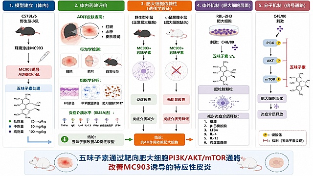
将提供的五味子素抗特应性皮炎机制图全部转换为全中文版,保留原有的全部模块布局、实验流程、结构式和配色不变,所有英文文字翻译为规范中文: 1. 模块标题改为:1. 模型建立(体内)、2. 体内药效评价、3. 肥大细胞依赖性(遗传学验证)、4. 体外机制(肥大细胞层面)、5. 分子机制(信号通路) 2. 所有标注文字翻译为中文:C57BL/6野生型小鼠、局部涂抹MC903、MC903诱导AD模型小鼠、五味子素处理、低剂量25 mg/kg、中剂量50 mg/kg、高剂量100 mg/kg;AD样皮肤损伤:红斑、水肿、炎症浸润;行为学检测:搔抓、擦耳、自发行为;组织学分析:HE染色、甲苯胺蓝染色、类胰蛋白酶染色;炎症介质水平(ELISA法):TSLP、IgE、IL-4、IL-13、组胺、类胰蛋白酶、LTB4;结论:五味子素改善AD炎症表型;野生型小鼠(正常肥大细胞)、c-kit敲除小鼠(肥大细胞缺失)、MC903+五味子素、皮损改善、炎症介质减少;无明显改善、炎症介质无降低;结论:抗AD作用依赖肥大细胞;RBL-2H3肥大细胞、刺激:C48/80、五味子素、抑制脱颗粒、减少炎症介质释放:组胺、β-己糖胺酶、LTB4、IL-4、IL-13、类胰蛋白酶;C48/80刺激、PI3K、AKT、mTOR、P代表磷酸化、抑制线代表五味子素抑制、肥大细胞活化、炎症介质释放;底部大标题改为:五味子素通过靶向肥大细胞并抑制PI3K/AKT/mTOR信号通路缓解AD炎症和瘙痒 保持正确的五味子素结构式完全不变,整体风格保持专业学术,适合中文论文使用。将提供的五味子素抗特应性皮炎机制图全部转换为全中文版,保留原有的全部模块布局、实验流程、结构式和配色不变,所有英文文字翻译为规范中文:
1. 模块标题改为:1. 模型建立(体内)、2. 体内药效评价、3. 肥大细胞依赖性(遗传学验证)、4. 体外机制(肥大细胞层面)、5. 分子机制(信号通路)
2. 所有标注文字翻译为中文:C57BL/6野生型小鼠、局部涂抹MC903、MC903诱导AD模型小鼠、五味子素处理、低剂量25 mg/kg、中剂量50 mg/kg、高剂量100 mg/kg;AD样皮肤损伤:红斑、水肿、炎症浸润;行为学检测:搔抓、擦耳、自发行为;组织学分析:HE染色、甲苯胺蓝染色、类胰蛋白酶染色;炎症介质水平(ELISA法):TSLP、IgE、IL-4、IL-13、组胺、类胰蛋白酶、LTB4;结论:五味子素改善AD炎症表型;野生型小鼠(正常肥大细胞)、c-kit敲除小鼠(肥大细胞缺失)、MC903+五味子素、皮损改善、炎症介质减少;无明显改善、炎症介质无降低;结论:抗AD作用依赖肥大细胞;RBL-2H3肥大细胞、刺激:C48/80、五味子素、抑制脱颗粒、减少炎症介质释放:组胺、β-己糖胺酶、LTB4、IL-4、IL-13、类胰蛋白酶;C48/80刺激、PI3K、AKT、mTOR、P代表磷酸化、抑制线代表五味子素抑制、肥大细胞活化、炎症介质释放;底部大标题改为:五味子素通过靶向肥大细胞并抑制PI3K/AKT/mTOR信号通路缓解AD炎症和瘙痒
保持正确的五味子素结构式完全不变,整体风格保持专业学术,适合中文论文使用。
[Main Image 2] Core ingredient analysis for anti-aging cream. [Atmosphere Style] Clean high-end scientific research medical style. [Image Quality] Super realistic 3D rendering, C4D product rendering style, Octane rendering, physical level rendering, high precision, clear picture. [Elements] In the center is the opened anti-aging cream jar, keep the product appearance (physical structure, material texture, color) absolutely consistent with the input product picture, eliminate any deformation and color difference. Around the product, six 3D molecular spheres representing different peptides are suspended, and a number of collagen molecular spheres representing five recombinant collagens surround the product. Each molecular sphere uses extremely thin lines to connect to the corresponding efficacy text description, showing 6 peptides anti-wrinkle matrix + five recombinant collagen nano-microspheres core technology. [Light and Shadow] Soft laboratory light, main light soft and even, auxiliary light bright molecular structure, transparent molecular light effect, clear layering. [Color Matching] Main color light bright off-white background, auxiliary color translucent molecular sphere soft light, dark green product bottle. [Perspective Composition] Centered composition, full screen no border, front view, molecular and product layering clear. [Text Layout] All text in English, core title "Time Reversal Key Multi-dimensional Collagen Regeneration" in large sans-serif font, small word descriptions for each component, font is high-end thin non-bold, text color dark green, high contrast with light background, text does not block the product subject. [Proportion Resolution] 1:1 proportion, 2K resolution, text clear and recognizable, super high-definition. [Negative prompt] Deformation, watermark, text confusion, product picture blurred, background messy, Chinese text, exaggerated text, extra decoration[Main Image 2] Core ingredient analysis for anti-aging cream. [Atmosphere Style] Clean high-end scientific research medical style. [Image Quality] Super realistic 3D rendering, C4D product rendering style, Octane rendering, physical level rendering, high precision, clear picture. [Elements] In the center is the opened anti-aging cream jar, keep the product appearance (physical structure, material texture, color) absolutely consistent with the input product picture, eliminate any deformation and color difference. Around the product, six 3D molecular spheres representing different peptides are suspended, and a number of collagen molecular spheres representing five recombinant collagens surround the product. Each molecular sphere uses extremely thin lines to connect to the corresponding efficacy text description, showing 6 peptides anti-wrinkle matrix + five recombinant collagen nano-microspheres core technology. [Light and Shadow] Soft laboratory light, main light soft and even, auxiliary light bright molecular structure, transparent molecular light effect, clear layering. [Color Matching] Main color light bright off-white background, auxiliary color translucent molecular sphere soft light, dark green product bottle. [Perspective Composition] Centered composition, full screen no border, front view, molecular and product layering clear. [Text Layout] All text in English, core title "Time Reversal Key Multi-dimensional Collagen Regeneration" in large sans-serif font, small word descriptions for each component, font is high-end thin non-bold, text color dark green, high contrast with light background, text does not block the product subject. [Proportion Resolution] 1:1 proportion, 2K resolution, text clear and recognizable, super high-definition. [Negative prompt] Deformation, watermark, text confusion, product picture blurred, background messy, Chinese text, exaggerated text, extra decoration
Ultra-detailed scientific illustration, flat vector style, showcasing the process of "Triparental Mating" for bacterial plasmid transfer. The image is divided into three clear stages from left to right. Stage 1: Three distinct bacterial cells in close proximity. 1 Donor E. coli: Contains a "mobilizable plasmid" with a visible "gene of interest" (like a fluorescent protein or antibiotic resistance marker) and an "oriT" site. 2 Helper E. coli: Contains a "conjugative plasmid" with visible "tra genes" (transfer genes) and pilus structures. 3 Recipient bacterium (e.g., a Rhizobium cell): Has a different cell wall shape, labeled, with no plasmid inside. Stage 2: The helper E. coli extends a "pilus" (a thin tube) to attach to the donor E. coli. The conjugative plasmid's tra genes mobilize the mobilizable plasmid. Show a clear arrow indicating the transfer of the mobilizable plasmid FROM the donor TO the helper cell. Stage 3: The helper E. coli, now carrying the mobilizable plasmid, extends a new pilus to attach to the recipient bacterium. Show a second arrow indicating the final transfer of the mobilizable plasmid INTO the recipient. The recipient bacterium now contains the plasmid. Use a distinct, consistent color scheme: Donor is Blue, Helper is Orange, Recipient is Green, Mobilizable Plasmid is Red, Conjugative Plasmid is Yellow. The background is clean white. All key elements have clear labels with leader lines. The style is educational, schematic, and extremely clear for a presentation slide.Ultra-detailed scientific illustration, flat vector style, showcasing the process of "Triparental Mating" for bacterial plasmid transfer. The image is divided into three clear stages from left to right. Stage 1: Three distinct bacterial cells in close proximity. 1 Donor E. coli: Contains a "mobilizable plasmid" with a visible "gene of interest" (like a fluorescent protein or antibiotic resistance marker) and an "oriT" site. 2 Helper E. coli: Contains a "conjugative plasmid" with visible "tra genes" (transfer genes) and pilus structures. 3 Recipient bacterium (e.g., a Rhizobium cell): Has a different cell wall shape, labeled, with no plasmid inside. Stage 2: The helper E. coli extends a "pilus" (a thin tube) to attach to the donor E. coli. The conjugative plasmid's tra genes mobilize the mobilizable plasmid. Show a clear arrow indicating the transfer of the mobilizable plasmid FROM the donor TO the helper cell. Stage 3: The helper E. coli, now carrying the mobilizable plasmid, extends a new pilus to attach to the recipient bacterium. Show a second arrow indicating the final transfer of the mobilizable plasmid INTO the recipient. The recipient bacterium now contains the plasmid. Use a distinct, consistent color scheme: Donor is Blue, Helper is Orange, Recipient is Green, Mobilizable Plasmid is Red, Conjugative Plasmid is Yellow. The background is clean white. All key elements have clear labels with leader lines. The style is educational, schematic, and extremely clear for a presentation slide.