创意预览图

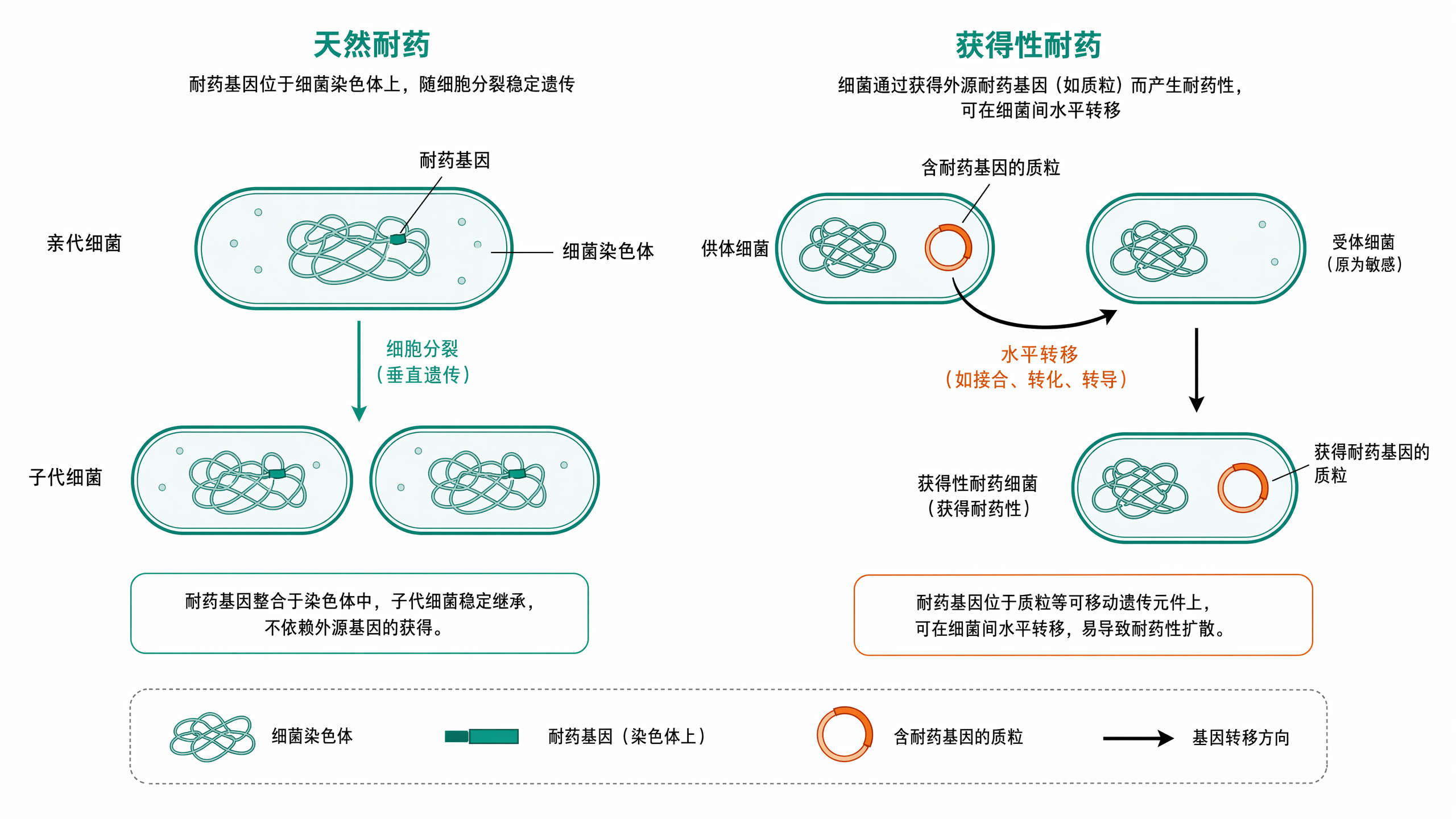
A scientific diagram comparing intrinsic resistance vs acquired resistance in bacteria, flat academi

Ultra-detailed scientific illustration, flat vector style, showcasing the process of "Triparental Mating" for bacterial plasmid transfer. The image is divided into three clear stages from left to right. Stage 1: Three distinct bacterial cells in close proximity. 1 Donor E. coli: Contains a "mobilizable plasmid" with a visible "gene of interest" (like a fluorescent protein or antibiotic resistance marker) and an "oriT" site. 2 Helper E. coli: Contains a "conjugative plasmid" with visible "tra genes" (transfer genes) and pilus structures. 3 Recipient bacterium (e.g., a Rhizobium cell): Has a different cell wall shape, labeled, with no plasmid inside. Stage 2: The helper E. coli extends a "pilus" (a thin tube) to attach to the donor E. coli. The conjugative plasmid's tra genes mobilize the mobilizable plasmid. Show a clear arrow indicating the transfer of the mobilizable plasmid FROM the donor TO the helper cell. Stage 3: The helper E. coli, now carrying the mobilizable plasmid, extends a new pilus to attach to the recipient bacterium. Show a second arrow indicating the final transfer of the mobilizable plasmid INTO the recipient. The recipient bacterium now contains the plasmid. Use a distinct, consistent color scheme: Donor is Blue, Helper is Orange, Recipient is Green, Mobilizable Plasmid is Red, Conjugative Plasmid is Yellow. The background is clean white. All key elements have clear labels with leader lines. The style is educational, schematic, and extremely clear for a presentation slide.Ultra-detailed scientific illustration, flat vector style, showcasing the process of "Triparental Mating" for bacterial plasmid transfer. The image is divided into three clear stages from left to right. Stage 1: Three distinct bacterial cells in close proximity. 1 Donor E. coli: Contains a "mobilizable plasmid" with a visible "gene of interest" (like a fluorescent protein or antibiotic resistance marker) and an "oriT" site. 2 Helper E. coli: Contains a "conjugative plasmid" with visible "tra genes" (transfer genes) and pilus structures. 3 Recipient bacterium (e.g., a Rhizobium cell): Has a different cell wall shape, labeled, with no plasmid inside. Stage 2: The helper E. coli extends a "pilus" (a thin tube) to attach to the donor E. coli. The conjugative plasmid's tra genes mobilize the mobilizable plasmid. Show a clear arrow indicating the transfer of the mobilizable plasmid FROM the donor TO the helper cell. Stage 3: The helper E. coli, now carrying the mobilizable plasmid, extends a new pilus to attach to the recipient bacterium. Show a second arrow indicating the final transfer of the mobilizable plasmid INTO the recipient. The recipient bacterium now contains the plasmid. Use a distinct, consistent color scheme: Donor is Blue, Helper is Orange, Recipient is Green, Mobilizable Plasmid is Red, Conjugative Plasmid is Yellow. The background is clean white. All key elements have clear labels with leader lines. The style is educational, schematic, and extremely clear for a presentation slide.

创建一幅科学准确的分子机制插图,主题为光合作用的光反应与暗反应过程(Photosynthesis: Light-dependent & Light-independent Reactions),采用高水平细胞生物学期刊风格(如Nature Plants风格),整体为横向分步机制图,清晰展示类囊体膜上的光反应如何驱动叶绿体基质中的暗反应(卡尔文循环)。左侧示意光反应阶段:展示类囊体膜结构,描绘光系统II(PSII)吸收光子激发电子,经电子传递链(含质体醌PQ、细胞色素b6f复合体、质体蓝素PC)传递至光系统I(PSI),电子最终还原NADP⁺为NADPH。同时展示水的光解(释放O₂)以及ATP合酶利用质子梯度(ΔpH)合成ATP的化学渗透过程。中间区域展示ATP与NADPH从类囊体膜释放,作为能量与还原力进入叶绿体基质。右侧展示暗反应阶段(卡尔文循环):描绘CO₂的固定(由Rubisco催化,生成3-磷酸甘油酸3-PGA)、还原(利用NADPH和ATP将3-PGA还原为3-磷酸甘油醛G3P/三碳糖)以及RuBP的再生过程。机制图中可醒目地标注“光反应”与“暗反应”两大板块,展示类囊体膜的磷脂双分层、光合色素(如叶绿素a/b)在PSII/PSI中的示意图,以及质子动力势驱动ATP合酶旋转催化的概念图。总体保持教材级概括性,逻辑主线清晰,避免过多调节分支,适合高中生物教学配图。整体图像要求结构准确、箭头逻辑清晰、颜色编码统一(如绿色调代表光合作用、蓝色箭头代表电子传递、红色箭头代表物质合成)。画面中需包含清晰可读的中文标题、中文过程标签与中文分子标注,例如“光反应”“暗反应”“类囊体膜”“叶绿体基质”“光系统II(PSII)”“光系统I(PSI)”“电子传递链”“ATP合酶”“NADPH”“卡尔文循环”“CO₂固定”“C₃还原”“RuBP再生”“Rubisco”等,所有中文说明排版规范,适合课堂教学展示与生物学科研汇报配图。创建一幅科学准确的分子机制插图,主题为光合作用的光反应与暗反应过程(Photosynthesis: Light-dependent & Light-independent Reactions),采用高水平细胞生物学期刊风格(如Nature Plants风格),整体为横向分步机制图,清晰展示类囊体膜上的光反应如何驱动叶绿体基质中的暗反应(卡尔文循环)。左侧示意光反应阶段:展示类囊体膜结构,描绘光系统II(PSII)吸收光子激发电子,经电子传递链(含质体醌PQ、细胞色素b6f复合体、质体蓝素PC)传递至光系统I(PSI),电子最终还原NADP⁺为NADPH。同时展示水的光解(释放O₂)以及ATP合酶利用质子梯度(ΔpH)合成ATP的化学渗透过程。中间区域展示ATP与NADPH从类囊体膜释放,作为能量与还原力进入叶绿体基质。右侧展示暗反应阶段(卡尔文循环):描绘CO₂的固定(由Rubisco催化,生成3-磷酸甘油酸3-PGA)、还原(利用NADPH和ATP将3-PGA还原为3-磷酸甘油醛G3P/三碳糖)以及RuBP的再生过程。机制图中可醒目地标注“光反应”与“暗反应”两大板块,展示类囊体膜的磷脂双分层、光合色素(如叶绿素a/b)在PSII/PSI中的示意图,以及质子动力势驱动ATP合酶旋转催化的概念图。总体保持教材级概括性,逻辑主线清晰,避免过多调节分支,适合高中生物教学配图。整体图像要求结构准确、箭头逻辑清晰、颜色编码统一(如绿色调代表光合作用、蓝色箭头代表电子传递、红色箭头代表物质合成)。画面中需包含清晰可读的中文标题、中文过程标签与中文分子标注,例如“光反应”“暗反应”“类囊体膜”“叶绿体基质”“光系统II(PSII)”“光系统I(PSI)”“电子传递链”“ATP合酶”“NADPH”“卡尔文循环”“CO₂固定”“C₃还原”“RuBP再生”“Rubisco”等,所有中文说明排版规范,适合课堂教学展示与生物学科研汇报配图。